Biomedical Imaging Systems
Hello, dear friend, you can consult us at any time if you have any questions, add WeChat: daixieit
Biomedical Imaging Systems
Magnetic Resonance Imaging (MRI) Project
In this project you will image simulated tissue phantom. The goal of this project is to understand and implement a spin-echo imaging sequence and to reconstruct an NMR image.
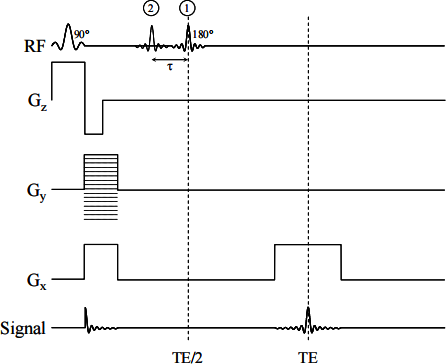
Consider the NMR imaging sequence shown.
To make things easier, we will assume a 2-dimensional spin distribution, so no selective excitation is necessary, and the spin densities are the same everywhere in the object. The object contains both water and fat with the following characteristics:
Water: T1 = 500 msec
T2 = 200 msec
γ0 = 42.57 MHz/Tesla
Fat: T1 = 300 msec
T2 = 100 msec
γ = γ0 + 3.5 ppm = (1+3.5·10-6)·γ0
Other necessary information:
The static magnetic field (B0) is along the Z-axis and of strength 1.5 Tesla
The time to echo (TE) is approximately 15 msec
The repetition time (TR) is approximately 500 msec
The duration of the readout is 8 msec
The duration of the 90-degree and 180-degree pulses is 62.5 µsec
B1 is strong enough so that both water and fat spins are fully rotated into the transverse plane with the same initial phase and the 180-degree pulse performs perfect phase inversion of both the water and fat components of the transverse magnetization.
The goal of the project is to perform the imaging sequence and to reconstruct images. Overall, you will develop a pulse sequence to image proton spins during readout for the 180 degree pulse at position 1 and position 2, and then, assuming that τ, the time between positions 1 and 2, is chosen such that 2/0 πτγτω =⋅⋅∆=⋅∆ B (hint, hint), develop a procedure to image only the water component or only the fat component using measurements obtained from positions 1 and 2.
To achieve this goal, you will write a time discretized simulation of the pulse sequence presented above, generate NMR signals, process them and, finally, produce two images separately highlighting fat and water spins. In addition, you will answer specific questions given below. To answer these questions, you will be supplied with:
A. 2-D array of mixed water and fat spins. Specifically, three 64 by 64 data matrices (corresponding to a 64 mm by 64 mm image): a matrix of gyromagnetic ratios, a matrix of spin-lattice relaxation times (T1) and a matrix of spin-spin relation times (T2), will be provided.
B. Skeleton of a Matlab script (nmr_template.m) for NMR Spin-Echo imaging where you should replace all the question marks and add some more commands. (Rename this file to nmr_YourLastName.m)
C. MATLAB routine (bloch.m) that will solve the time discretized Bloch equations for you (note that this function assumes 1.5 T field, i.e., big gorilla is always on because of this function !!!).
D. Matlab function called recover.m which solves the Bloch equations for long periods of longitudinal relaxation (again, 1.5 T field is on here since recover.m uses bloch.m).
Rules of the Game:
You should work alone on the project and submit (via canvas) a written report (PDF only, no zip files, or any other file types).
Prepare a report by answering the questions as requested (see below). Your report will have graphs and images. You will also submit (via canvas) your final “.m” file. In the report, please write a few sentences about what you did, equation(s) you utilized, and comment on the appearance of the images. You could also describe your frustrations. In any case, say something interesting!!!
The partnering is allowed. If you team up with anyone, use it as an environment where you can discuss questions, etc. – you should not split up the problems and then share solutions. I expect you to do your own work, but when you get stuck, please ask myself or classmates for help. Therefore, getting help from anyone other than your teammates (students in the class) or the instructor (i.e., me) or copying solutions from anyone will be considered a violation of the rules.
Name your plots and images, and label the axes.
If you are having problems, please come and see me, or initiate a discussion on canvas, or send me an e-mail.
Please, answer the following questions
1a) Develop a pulse sequence to image proton spins during readout for the 180 degree pulse at position 1.
Produce Fourier image and m(x,y) image (2 figures, 15 points)
1b) Develop a pulse sequence to image proton spins during readout for the 180 degree pulse at position 2.
Produce Fourier image and m(x,y) image (2 figures, 15 points)
2) Describe a procedure to image only the water component or only the fat component using measurements obtained in question 1. Produce two images to demonstrate your answer. Justify your answer by a diagram or, even better, signals from your simulations. Describe your results using vector model (at least 2 figures with images, 30 points)
3) After you produced two images (question 1) to reconstruct the fat-only and water-only images (question 2), are the images perfect? Explain the image artifacts, if any, or explain why there are no artifacts. Here you may want to use NMR signal for a single fat spin and compare it to the NMR signal from a single water spin (use your code from questions 1-2 to produce signals and images). Are the signals the same? Why or why not? How about the reconstructed images: are they the same? Why or why not? (20 points)
4) What assumptions did we use to make the simulation and how would this experiment change in a real NMR system? How would you change the simulation to include a distribution of spin densities? How would you change the simulation to get T1 weighted images? T2 weighted images? (20 points)
Hints: For question 1, recall/examine how fft.m and fftshift.m work in MATLAB. For question 2, use vector model to understand what is happening with water spin versus fat spin during MRI sequence. Why the specific delay was introduced in the sequence above? If you can understand why the magnetizations and signal are what they are for a single spin, then imaging water only or fat only will be easy.
Good luck!
2023-09-18
Magnetic Resonance Imaging (MRI) Project